Seeing Life Up Close: The Astonishing Image Of The Cell
Have you ever stopped to think about what makes up all living things, from the smallest blade of grass to the tallest tree, or even you? It's pretty amazing, isn't it? Well, the answer lies in something incredibly tiny, something you cannot see with just your eyes: the cell. Getting a good image of the cell lets us peek into this hidden world, revealing the fundamental building blocks of all life around us.
For a long time, these tiny structures were a complete mystery. People just knew that living things grew and changed, but the how was a big question. Then, with the invention of the microscope, a whole new universe opened up. Suddenly, we could see these little compartments, and the idea of the cell came to be. It was, you know, a really big step in science.
Today, finding the right image of the cell is easier than ever, thanks to tools that offer the most comprehensive image search on the web. You can see a wide range of pictures, from basic diagrams to incredibly detailed photos taken with advanced equipment. It is that simple to explore the tiny bits that make us who we are.
- Lionel Richie And Diana Ross
- Harmony Ether Leaks
- City Of Edmond
- One Piece English Dub
- Blacksburg Va Weather
Table of Contents
- What is an Image of the Cell?
- How We Get to See Them: Microscopy Methods
- Different Cells, Different Looks
- The Beauty and Wonder of Cell Pictures
- Common Questions About Cell Images
- Finding and Appreciating Cell Images Today
What is an Image of the Cell?
An image of the cell is, quite simply, a visual record of these tiny life units. These pictures help us see what cells look like, how they are put together, and what little parts they have inside. They are, you know, like maps of the very smallest bits of life.
These images can come in many forms. Some are drawings or diagrams that help explain complex ideas. Others are actual photographs taken through powerful instruments. They show us everything from a cell's outer boundary to the tiny structures working away inside it.
It's pretty amazing to think about how far we have come in getting these pictures. Early images were very simple, just showing basic shapes. Now, we can see incredible detail, almost like walking inside a cell. This progress really helps us learn a lot more.
- Uruguay National Football Team Vs Argentina National Football Team Lineups
- Weather Newburgh Ny
- Weather Troy Mi
- Dave Campbell Texas Football
- Saudi Council Of Engineers
Why These Pictures Matter
Getting a clear image of the cell is incredibly important for many reasons. For one thing, it helps us teach and learn about biology. It is much easier to grasp how something works when you can actually see it. Textbooks and lectures come alive with good pictures.
These images are also vital for scientific study. Researchers use them to study diseases, like cancer, by looking at how sick cells are different from healthy ones. They help scientists understand how medicines might affect cells, too. So, in some respects, they guide discovery.
Furthermore, an image of the cell helps us appreciate the complexity of life. It shows us that even the smallest parts of living things are incredibly organized and busy. It gives us a better sense of how everything fits together, which is rather neat.
How We Get to See Them: Microscopy Methods
You might wonder how we get such clear pictures of something so small. It's not like taking a picture with your phone, you know. Special tools called microscopes are needed. These tools use different ways to make tiny things appear much bigger. Basically, they let us zoom in a lot.
Over time, the ways we look at cells have changed a lot. Early microscopes were very basic. Now, we have highly advanced machines that can show us things once thought impossible to see. This progress just keeps going, which is pretty cool.
Each type of microscope offers a unique way to view cells. Some are good for seeing living cells in action. Others are better for showing really fine details of cell parts. It really depends on what you want to learn from the image of the cell.
Light Microscopes: The First Look
Light microscopes are probably what most people think of when they hear the word "microscope." These tools use light to make things bigger. You put a very thin slice of something on a glass slide, and light shines through it. Lenses then bend the light to make the image appear larger. This is how cells were first seen, you know.
They are great for seeing general cell shapes and sizes. You can often see the main parts, like the nucleus in many cells. Many school labs use these types of microscopes because they are pretty straightforward to use. They give you a good overall image of the cell.
Sometimes, scientists add special colors or dyes to the cells. This makes certain parts stand out more clearly under the light microscope. It helps them spot specific structures, which is quite helpful for study.
Electron Microscopes: Seeing in Fine Detail
When you need to see really, really tiny parts inside a cell, electron microscopes come into play. Instead of light, these machines use a beam of electrons. Electrons are much smaller than light waves, so they can show much finer details. This allows for an incredibly detailed image of the cell.
There are a couple of main kinds of electron microscopes. A transmission electron microscope, or TEM, shoots electrons through a very thin sample. It gives you a flat, black-and-white picture of the inside of a cell. It shows structures that are too small for light microscopes to pick up.
Then there is the scanning electron microscope, or SEM. This one bounces electrons off the surface of a sample. It creates amazing 3D-like images of the cell's outer surface. You can see bumps and grooves, which is really cool to look at. Both types offer unique perspectives.
Newer Ways to Look: Confocal and Super-Resolution
Science keeps moving forward, and so do the ways we get an image of the cell. Confocal microscopes are a more modern type of light microscope. They use lasers to scan the cell point by point. This helps remove blurry light from above and below the focus area. You get much sharper, clearer images, sometimes in color, which is pretty neat.
Super-resolution microscopy is even newer. It breaks the old limits of light microscopes, letting us see things even smaller than what was once thought possible with light. It is almost like magic, allowing scientists to watch tiny cell parts move and interact in real time. This really helps us understand cell processes.
These advanced methods let scientists ask new questions about how cells work. They provide incredibly precise images, showing how molecules behave within a cell. This helps with everything from basic research to finding new ways to treat illnesses. It's truly exciting stuff, you know.
Different Cells, Different Looks
Not all cells look the same, even though they all share some basic features. Just like houses, they have different shapes and sizes depending on their job. An image of the cell can show us these wonderful differences. It's really quite varied.
For example, a nerve cell looks very different from a skin cell. This is because they have very different tasks to do in the body. Seeing these distinct forms helps us understand their functions better. It just makes sense, doesn't it?
Let's take a quick look at some common types of cells and what their images might show us. This will give you a better idea of the variety out there. There's so much to explore, really.
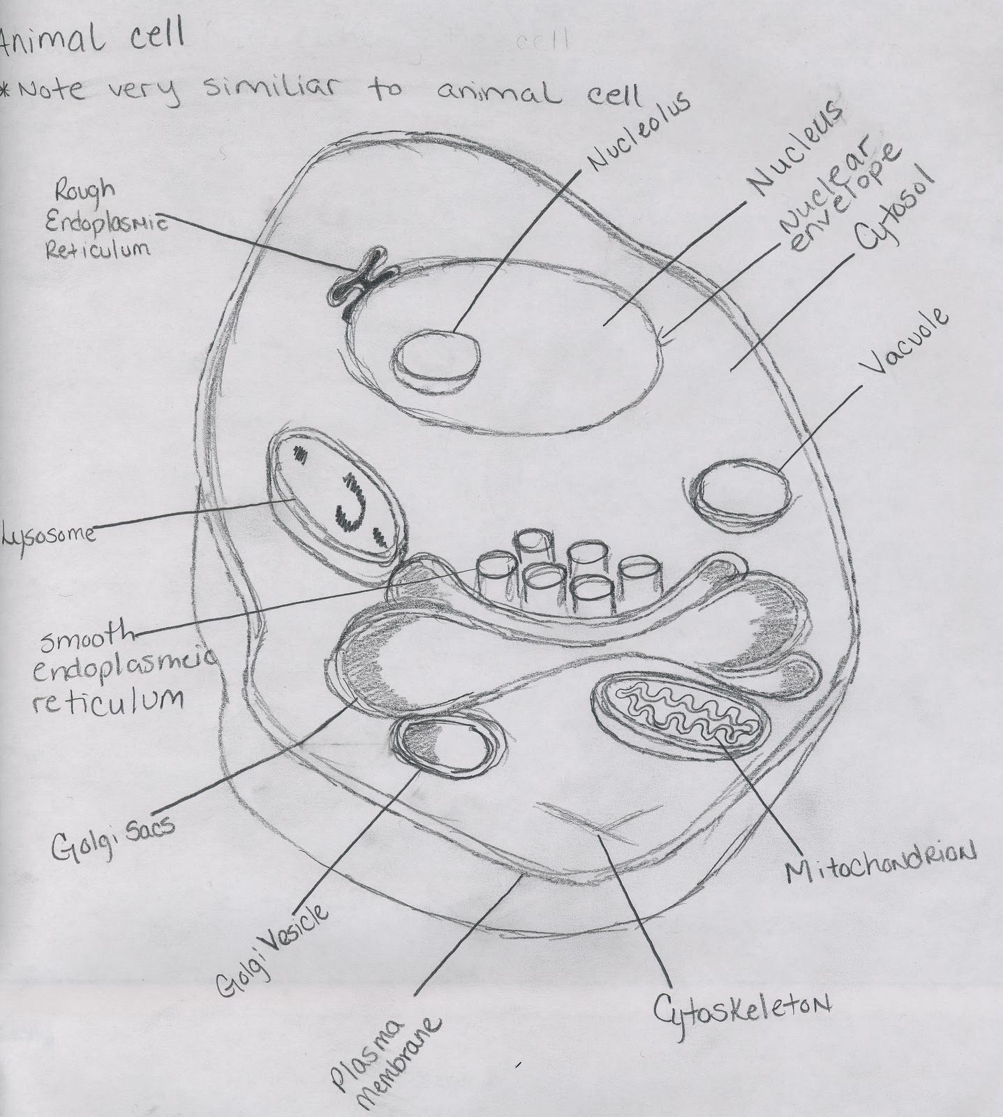
Animal Cells: Our Own Building Blocks
Animal cells, like the ones that make up your body, typically have an irregular, somewhat rounded shape. They do not have a stiff outer wall like plant cells. This allows them to be more flexible, which is helpful for many body functions. You can often see the nucleus, which is like the cell's control center, very clearly.
An image of the cell from an animal might show you tiny powerhouses called mitochondria. These make energy for the cell. You might also spot other small sacs and tubes that help with different jobs, like moving things around or making proteins. It's a busy little world inside.
Human blood cells, for instance, are a great example. Red blood cells are shaped like tiny donuts without a hole, which helps them carry oxygen. White blood cells have more varied shapes as they move around to fight off bad stuff. They are quite distinct, you know.
Plant Cells: The Green World Up Close
Plant cells have a very different appearance from animal cells. They usually have a more fixed, box-like shape. This is because they have a strong cell wall outside their cell membrane. This wall gives plants their stiffness and support, which is pretty important for standing tall.
An image of the cell from a plant will often show you big, green structures called chloroplasts. These are where plants make their food using sunlight, a process called photosynthesis. They are what make plants green, actually.
You will also likely see a large central vacuole in a plant cell. This is a big sac that stores water and helps keep the cell firm. When a plant wilts, it is often because these vacuoles are losing water. So, they are quite vital for plant health.
Bacteria and Other Tiny Life Forms
Bacteria are some of the smallest and simplest cells around. They are called prokaryotic cells, meaning they do not have a nucleus or other complex parts enclosed in membranes. An image of the cell for bacteria will show a much simpler internal structure compared to plant or animal cells. They are, in a way, very basic.
Their shapes can vary a lot, though. Some are rod-shaped, others are round, and some are spiral. Many have little tails called flagella that help them move around. Seeing these shapes helps scientists identify different types of bacteria, which is very important for medicine and other fields.
Other tiny life forms, like fungi and algae, also have unique cell structures. Fungal cells often have cell walls, but they are made of different materials than plant cell walls. Algae cells might have chloroplasts like plants, but they can be very diverse in form. Each one offers a fascinating image of the cell.
The Beauty and Wonder of Cell Pictures
Beyond their scientific value, an image of the cell can be truly beautiful. When you look at them, especially those taken with advanced techniques and colored in, they can look like abstract art. The patterns, the colors, the intricate details – they are really quite stunning.
These pictures remind us that even at the smallest scales, life is full of wonder and complexity. It is pretty humbling to think that every living thing, including us, is made of these tiny, busy units. They are, you know, the silent workers that keep everything going.
Many artists and photographers are inspired by these microscopic views. They show us that science and art can truly meet. It encourages us to look closer at the world around us, even the parts we cannot easily see. It just shows how much there is to appreciate.
Common Questions About Cell Images
People often have questions when they first see an image of the cell. It's natural to be curious about something so small and fundamental. Here are a few common things people ask, which is pretty typical.
1. Why do cell images sometimes have different colors?
Often, cells are naturally clear or transparent. To make different parts stand out, scientists use special dyes or stains. These chemicals stick to specific parts of the cell, giving them color. This helps researchers see and study them better. Sometimes, colors are also added digitally to highlight different structures in complex images, which is quite useful.
2. Can we see living cells in real-time?
Yes, absolutely! While some imaging methods require cells to be prepared in ways that mean they are no longer living, many modern microscopes allow us to observe living cells. Techniques like phase-contrast microscopy or confocal microscopy let scientists watch cells move, divide, and interact in real-time. This is incredibly valuable for understanding dynamic biological processes, you know.
3. How small is the smallest cell we can image?
The smallest cells are typically bacteria, and some are incredibly tiny, just a few hundred nanometers across. A nanometer is one billionth of a meter! With electron microscopes and super-resolution light microscopes, we can image these tiny cells and even see some of their internal parts. We can even see large molecules, which is pretty amazing when you think about it.
Finding and Appreciating Cell Images Today
Thanks to the internet, finding an image of the cell is easier than it has ever been. Tools like Google Image Search provide "the most comprehensive image search on the web," offering a vast collection of pictures from various sources. You can find everything from simple diagrams to stunning, high-resolution photographs. It's a great way to explore this topic further.
When you look at these images, take a moment to really think about what you are seeing. Consider the incredible technology that made the picture possible. Think about the tiny, complex world it reveals. It is, you know, a window into the very essence of life.
We encourage you to explore these fascinating visuals. Learn more about cells on our site, and perhaps even link to this page for more detailed scientific explanations. The more you look, the more you will appreciate the intricate beauty that makes up everything around us. It's truly a rewarding experience, actually.
- Elly Clutch Porn
- Weather Buffalo Grove Il
- Mashable Connections Today
- Clear Lake Iowa
- Josh Hutcherson Naked
Cell Types and Structure: Animal Cell
Cell - Original Art | Florian Bertmer
Amazing Cell Coloring - Coloring Page

